Ultra Wideband-Based Imaging Technology for Stroke Detection
- contact:
- project group:
Medical measurement technology, Numerical field calculation, Imaging
- Partner:
Institute for High-Frequency-Technology and Electronics (IHE)
Stroke is still one of the main reasons for disability and death in developed nations. For patients with stroke it is extremely important to receive the right treatment as fast as possible because the tissue surrounding the stroke core immediately starts to become necrotic.  Unfortunately, a treatment can only begin if the cause of a stroke is verified, as there are two different types of stroke with exactly the same symptoms. For each cause completely different medication is needed. Ischemic strokes caused by an occlusion of a vessel e.g. by a clot occurs most often. In this case a medication with hemodilution is needed. On the contrary a rupture of a vessel induces the hemorrhagic stroke and a medication promoting coagulation is needed. For the diagnosis two different procedures are applied today: the one is the computed tomography (CT), the other is the magnetic resonance imaging (MRI). Unfortunately, both procedures often lead to a long time span until therapy can be initiated. These systems are very expensive so that not every hospital can operate a CT or MRI, and these systems require professional personnel not always available for 24h per day and 7 days per week.
The aim of this project is to develop a new diagnostic device based on ultra wideband techniques (UWB). Such a device will not need big and expensive hardware and thus could even be used in ambulances.
A finite-difference time-domain method (FDTD) simulation environment with a model of a human head will be used to develop the necessary algorithms. The software SEMCAD X 14.8 distributed by Schmid & Partner Engineering AG is used for the simulation environment.
It’s a joint project by the Institute for High-Frequency-Technology and Electronics (IHE) and the In-stitute of Biomedical Engineering (IBT). The construction of the antennas and the other hardware are mainly performed by the IHE, the development of the algorithms is done in cooperation and the phantom as well as the computer model in SEMCAD is performed by the IBT.
For the construction of the model there are different things to take in to account:
- The permittivity of human tissue is a complex function of frequency. A good description for it is a 4-pole Cole & Cole equation.
- Which Frequency range can be used? An important point with respect to the spatial resolu-tion and the computing duration in FDTD.
- There is no or not enough information about the electromagnetic properties of tissue af-fected by stroke.
How the stroke diagnosis should work:
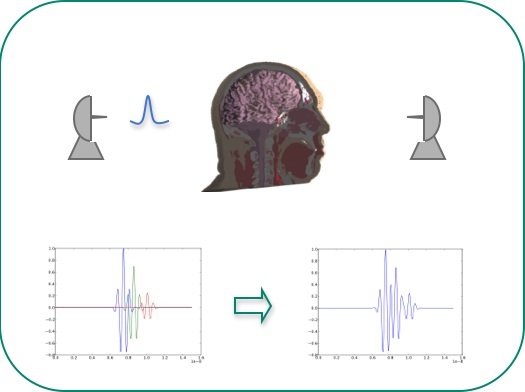
One or a set of antennas sends a short electromagnetic wave through the object to be inspected. The reflected and transmitted signals contain information about the electromagnetic properties, which the algorithm extracts. That information can then be used to reconstruct e.g. an image of the insides.
However, the following problems have to be solved:
- One of the problems is caused by the reflections of the skin and the skull which prevents us from sending and receiving a big enough amount of energy to or from the inside tissues.
- Another problem limits the frequency range and therefore the spatial resolution. Its cause is the conductivity of biological tissues. With growing frequencies the conductivity increases too. This means that no high frequent component of a signal is able to get through.
- A third problem interrelated with the problem described above is the duration of the signal with respect to the speed of light. The duration of propagation is shorter than the signal’s duration. This leads to a convoluted signal consisting of numerous reflections. A so-called deconvolution is necessary.

